Research Type
ResearcherPerson
Tori ZiegerNotes
Answers for the osteology program:
Sex:
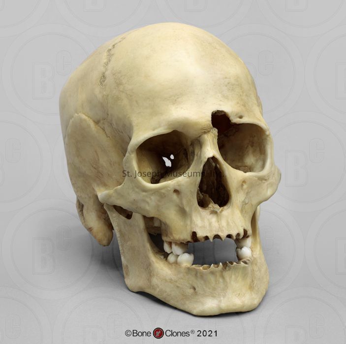
Male. The supraorbital ridge and external occipital protuberance/nuchal crest are well developed. The mastoid process is robust and the supraorbital ridge is rounded. The mental eminence on the mandible is well defined.
Age:
Adult. Third molars have not or have barely begun to erupt.This puts the age of the individual between 17-21. Sutures are closed but very visible. Age would be estimated as a young adult between the ages of 17-25.
Trauma:
Perimortem projectile trauma.
There are 12 remaining teeth. RIGHT MANDIBLE: first, second and third molar. RIGHT MAXILLA: first, second and third molar. LEFT MANDIBLE: first, second, and third molar, second premolar. LEFT MAXILLA: first and second molar. The third molars are visible but not erupted. All teeth that are missing were lost after death; the tooth sockets do not show signs of healing like they would if they were lost during life. The tooth surfaces are all covered with silver fillings. The first molar (left mandible) has its distal buccal surfaces chipped, with some chipping on the distal side of the second premolar (left mandible).
On the left frontal bone, right above the left orbit (right above the eye socket), there is a semicircular entry point for a bullet. The side indicates it is a small to medium caliber. No significant radiating fractures from the point of entry, however there is damage to the superior orbital wall of the frontal bone (the top part of the eye socket) and the sphenoid caused by the gunshot.
The exit wound is on the occipital bone, right on the lambdoid suture. A large radiating fracture travels from the exit wound to the squamosal suture. The beveling on the outside of the bone shows this is the exit wound. The entry and exit wound line up almost completely, indicating that the trajectory was a straight line from the forehead to the back of the head. Without additional details, it is impossible to confidently address if this was self inflicted or caused by another person, however it is harder to get a straight trajectory with a self-inflicted gunshot wound.