Research Type
ResearcherPerson
Tori ZiegerNotes
Answers for the osteology program:
Sex:
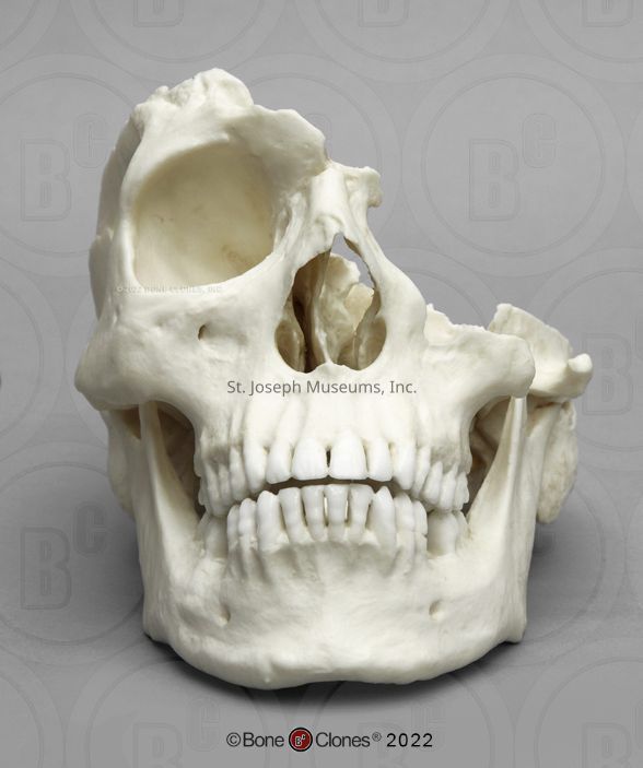
Male. The external occipital protuberance/nuchal crest (what is visible) is prominent. The mastoid processes are large. The mandible is more square shaped. The supraorbital margins are rounded.
Age:
Adult, likely 25+. What is visible of the lambdoid suture is fused but incomplete closure. The squamosal suture that is visible is open, but it is to be noted that severe trauma can interrupt cranial sutures and the lack of closure could be a result of the trauma. All dentition that is present is adult. The third molars are fully erupted
Trauma:
Perimortem multiple sharp force traumas and an antemortem healed trauma on the nose.
There are 30 teeth remaining, 16 in the maxilla (all of them) and 14 in the mandible. The mandible is missing both right and left first molars, which were lost antemortem as evidenced by the healed socket. On the maxillary right second premolar there is a defect on the lingual side which indicates a cavity (called caries). On the maxillary left first premolar there is part of the enamel missing on the lingual surface that might have been chipped off near the time of death. On the mandibular right second molar there is a large cavity caused by caries on the buccal/occlusal surface. The mandibular left second premolar shows evidence of a cavity near the root line on the buccal surface. The mandibular left third molar also shows evidence of a cavity on the buccal/occlusal surface.
There is a healed trauma on the right nasal bones. The vomer (the bone in the middle of the nasal cavity) is displaced to the right. These are injuries indicative of a (healed) broken nose.
The most significant evidence of trauma is that the left side of the skull and a large portion of the right is completely missing.
Trauma A is to the left temporal bone, right behind the external auditory meatus and extending above the mastoid process. The bottom edge of this trauma is chipped. This injury matches a sharp trauma on the coronoid process and mandibular condyle (the top of the left side of the mandible). These injuries lineup when the jaw is placed in anatomical position.
Trauma B is to the left side of the occipital bone (posterior to the first trauma).
Trauma C is in the center of the occipital bone, through the cruciform evidence of the occipital bone (the projection on the inside where it looks like four lines meet). Secondary fractures extend to the foramen magnum and through the right side of the occipital, where the secondary fractures terminate in what could be a smaller Trauma D.
Trauma A and C are both angled to the right and angled down, meaning someone likely chopped the skull from above and to the left. Trauma B is more horizontal but still is angled downward, supporting the claim that someone was chopping from above.
Trauma E is on the right parietal. The termination of this trauma is all that is visible - most of the impact site is on the missing bone. The fracture caused by this injury extends anteriorly through the temporal bone and terminates at the squamosal suture.
Looking on the inside of the skull, there is a small Trauma F on the petrous portion of the left temporal bone (find the foramen magnum; the chunky bit above and to the side is the petrous portion. It's the inside of the ear canal). There’s a small Trauma G right behind the missing left eye socket. Both of these traumas have radiating fractures. There is evidence of a blunt trauma on the left eye socket. This could be associated with Trauma G.
It is likely there were more traumas but the impact site is missing with the missing bones. The straight edges on the right parietal bones, plus the chipping above the right eye socket, indicates most strongly the existence of other traumatic injuries, as well as the straight edge on the left temporal/parietal and the internal beveling there.
It is not possible to determine the order of strikes besides saying that Trauma A occurred before Trauma B.