Research Type
ResearcherPerson
Tori ZiegerNotes
Answers for osteology program:
Sex:
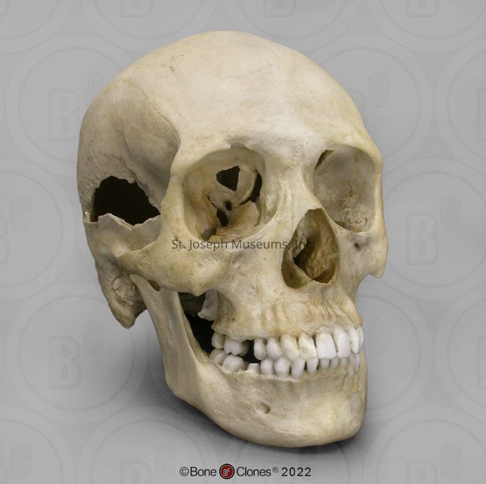
Female. External occipital protuberance/nuchal crest and mastoid processes are small. Supraorbital ridge is small and margin is sharp. The mental eminence is slightly prominent, but the mandible as a whole is small and rounded.
Age:
Older adult, likely over 50 years old. Teeth are all adult and the third molar is completely erupted. Sutures are fully closed with some aspects of all sutures having disappeared completely, indicating several years past peak closure.
Trauma:
Perimortem projectile trauma and antemortem fracture of the left styloid process. Small caliber
There are 28 teeth present, 14 in the mandible and 14 in the maxilla. MANDIBLE: left third molar is missing, but the socket is unhealed meaning the tooth was lost after death. The left first molar is missing with the socket healed, meaning it was lost during life. There is some gum recession visible, especially around the incisors and canines. The right first molar is almost completely missing due to cavities. The soft edges indicate this was not tooth loss due to a trauma. MAXILLA: the left first molar is missing with the socket healed, meaning the tooth was lost during life. The right first molar is missing but the socket is unhealed, meaning the tooth was lost after death. Significant gum recession particularly around incisors. There is evidence of gum disease around the molars, as seen with the atypical and bubbled appearance of where the gums would be.
There is a healed (or healing) fracture of the left styloid process (the thin projection behind the mastoid process) as evidenced by the thickening bone at the end of the fracture.
There are three small caliber projectile wounds on the left occipital bone. One stands alone and the figure 8 shape is caused by two bullets entering the skull. The small caliber is indicated by the general lack of destruction near the entrance site.
The exit wound(s) are not visible. It is likely that due to the small caliber of the bullets that there is no exit wound. The obvious evidence of trauma to the right temporal bone was caused by a “blow out” effect, which is caused by the rapid downward displacement of the brain. Two more blow out fractures can be seen in both eye sockets; the orbital shelf of both eyes fractured and were lost as a result of sudden pressure inside the skull due to the gunshot.