Research Type
ResearcherPerson
Tori ZiegerNotes
Answers for osteology program:
Sex:
Female. Supraorbital ridge is flat and supraorbital margins are sharp. Mastoid processes are small and external occipital protuberance/nuchal crest is small.
Age:
Young adult, likely younger than 30. Third molars are missing but the cavity is developed and unhealed, meaning they were lost during life. If third molars were unerupted, they are unlikely to be missing if other molars are not. Cranial sutures are fusing but are not closed completely.
Trauma:
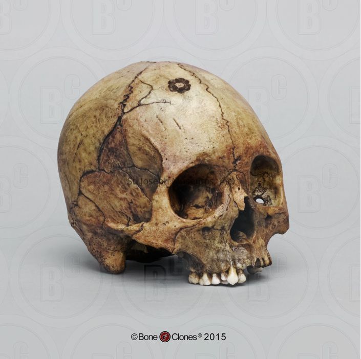
Perimortem incomplete trephination. Antemortem fracture. Perimortem radiating fractures
There are seven maxillary teeth present: right - canine, first and second premolar, first and second molar. Left - second molar. All teeth show evidence of being lost after death (unhealed sockets) with the exception of the left first molar and possibly the left third molar. The right first premolar shows a break on the lingual aspect of the enamel. Molars and second premolar on the right side show a degree of wear. The socket for the first molar on the left side shows signs of healing, though it is not complete. The holes for the roots are very shallow and the bone around the first molar has been damaged/broken off. This is likely done during life as postmortem breaks are unlikely to be as localized. The gum absorption seen around the left second molar in conjunction with the absorption seen here indicates gum disease. The lingual root for right first molar is slightly visible, indicating the beginning of the loss of bone around that tooth as well.
This individual has an unfused metopic suture. In infancy, the two halves of the frontal bone usually fuse completely, leaving no trace of that suture. In some individuals, that suture stays visible. This is a non-metric trait that would not have affected the life of the individual.
There is a healed fracture to the left nasal/maxilla, seen in the slight bowing/build up of bone into the left eye socket and the slightly irregular texture right under the corner of the eye socket. The vomer, the bone in the middle of the nasal cavity, is also slightly displaced to the left, likely because of the bony projection on the right.
Part of the left zygomatic arch is missing, but it is not possible to tell if that was done peri or post mortem.
The lambdoid suture is full of wormian bones, which are just extra bones in the skull. These are a non-metric trait and would not have affected the life of the individual.
On the frontal, right parietal, and right temporal bones there are several radiating fractures. These show minimal signs of healing, although the casting process could have affected the signs of healing. These radiating fractures do not have an obvious point of impact, but it is not caused by the incomplete trephination. It is possible that the trephination was in response to the injury that caused these fractures. The fractures radiate from the parietal towards the frontal bone and anterior of the skull. This indicates that the point of origin for the radiating fractures is likely on the parietal or temporal bones (as opposed to an injury to the face, for example).
The trephination is on the right frontal. The trephination was done with some sort of drill-like tool and did not perforate the skull completely. Usually these surgeries were done with a knife or other sharp object. There are 8 sites of drilling in a circular pattern. The site does not show signs of healing, although again the casting process could have obscured some of the signs. It is likely that this surgery and the causing trauma was the cause of death.